NCERT Solutions for Class 11 Biology chapter 21 Neural Control and Coordination PDF - eSaral

NCERT Solutions for Class 11 Biology chapter 21 Neural Control and Coordination PDF
Hey, are you a class 11 student and looking for ways to download NCERT Solutions for Class 11 Biology chapter 21 Neural Control and Coordination PDF? If yes. Then read this post till the end.In this article, we have listed NCERT Solutions for Class 11 Biology chapter 21 Neural Control and Coordination in PDF that are prepared by Kota’s top Doctor’s Faculties by keeping Simplicity in mind.
If you want to learn and understand class 11 Biology chapter 21 "Neural Control and Coordination" in an easy way then you can use these solutions PDF.
NCERT Solutions helps students to Practice important concepts of subjects easily. Class 11 Biology solutions provide detailed explanations of all the NCERT questions that students can use to clear their doubts instantly.
If you want to score high in your class 11 Biology Exam then it is very important for you to have a good knowledge of all the important topics, so to learn and practice those topics you can use eSaral NCERT Solutions.
In this article, we have listed NCERT Solutions for Class 11 Biology chapter 21 Neural Control and Coordination PDF that you can download to start your preparations anytime.
So, without wasting more time Let’s start.
Download NCERT Solutions for Class 11 Biology chapter 21 Neural Control and Coordination PDF
Question 1: Briefly describe the structure of the following:
1. Brain (b) Eye (c) Ear
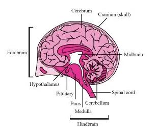
Solution. (A) Brain: Brain is the main coordinating centre of the body. It is a part of nervous system that controls and monitors every organ of the body. It is well protected by cranial meninges that are made up of an outer layer called dura mater, a thin middle layer called arachnoid, and an inner layer called pia mater.
It is divided into three regions − forebrain, midbrain, and hindbrain.
Forebrain: It is the main thinking part of the brain. It consists of cerebrum, thalamus, and hypothalamus.
(a) Cerebrum:
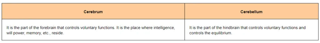
Cerebrum is the largest part of the brain and constitutes about four-fifth of its weight. Cerebrum is divided into two cerebral hemispheres by a deep longitudinal cerebral fissure. These hemispheres are joined by a tract of nerve fibre known as corpus callosum. The cerebral hemispheres are covered by a layer of cells known as cerebral cortex or grey matter. Cerebrum has sensory regions known as association areas that receive sensory impulses from various receptors as well as from motor regions that control the movement of various muscles. The innermost part of cerebrum gives an opaque white appearance to the layer and is known as the white matter.
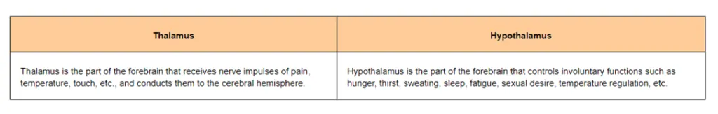
(b) Thalamus:
Thalamus is the main centre of coordination for sensory and motor signalling. It is wrapped by cerebrum.
(c) Hypothalamus:
It lies at the base of thalamus and contains a number of centres that regulate body temperature and the urge for eating and drinking. Some regions of cerebrum, along with hypothalamus, are involved in the regulation of sexual behaviour and expression of emotional reactions such as excitement, pleasure, fear, etc.
Midbrain:
It is located between the thalamus region of the forebrain and pons region of hindbrain. The dorsal surface of midbrain consists of superior and inferior corpora bigemina and four round lobes called corpora quadrigemina. A canal known as cerebral aqueduct passes through the midbrain. Midbrain is concerned with the sense of sight and hearing.
Hindbrain:
It consists of three regions − pons, cerebellum, and medulla oblongata.
(a) Pons is a band of nerve fibre that lies between medulla oblongata and midbrain. It connects the lateral parts of cerebellar hemisphere together.
(b) Cerebellum is a large and well developed part of hindbrain. It is located below the posterior sides of cerebral hemispheres and above medulla oblongata. It is responsible for maintaining posture and equilibrium of the body.
(c) Medulla oblongata is the posterior and simplest part of the brain. It is located beneath the cerebellum. Its lower end extends in the form of spinal cord and leaves the skull through foramen magnum.
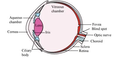
(B) Eye: Eyes are spherical structures that consist of three layers.
(a) The outer layer is composed of sclera and cornea.
(i) Sclera is an opaque tissue that is usually known as white of the eye. It is composed of a dense connective tissue.
(ii) Cornea is a transparent anterior portion of eye that lacks blood vessels and is nourished by lymph from the nearby area. It is slightly bulged forward and helps in focusing light rays with the help of lens.
(b) The middle layer of eye is vascular in nature and contains choroid, ciliary body, and iris.
(i) Choroid lies next to the sclera and contains numerous blood vessels that provide nutrients and oxygen to the retina and other tissues.
(ii) Ciliary body: The choroid layer is thin over posterior region and gets thickened in the anterior portion to form ciliary body. It contains blood vessels, ciliary muscles, and ciliary processes.
(iii) Iris: At the junction of sclera and cornea, the ciliary body continues forward to form thin coloured partition called iris. It is the visible coloured portion of eye.
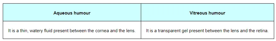
The eye contains a transparent, biconvex, and elastic structure just behind the iris. It is known as lens. The lens is held in position by suspensory ligaments attached to the ciliary body. The lens divides the eye ball into two chambers – an anterior aqueous and posterior vitreous chamber.
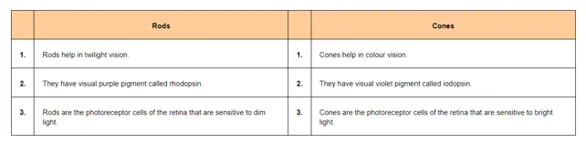
(c) The innermost nervous coat of eye contains retina. Retina is the innermost layer. It contains three layers of cells – inner ganglion cells, middle bipolar cells, and outermost photoreceptor cells. The receptor cells present in the retina are of two types – rod cells and cone cells.
(a) Rod cells –The rods contain the rhodopsin pigment (visual purple) that is highly sensitive to dim light. It is responsible for twilight vision.
(b) Cone cells –The cones contain the iodopsin pigment (visual violet) and are highly sensitive to high intensity light. They are responsible for daylight and colour visions.
The innermost ganglionic cells give rise to optic nerve fibre that forms optic nerve in each eye and is connected with the brain.
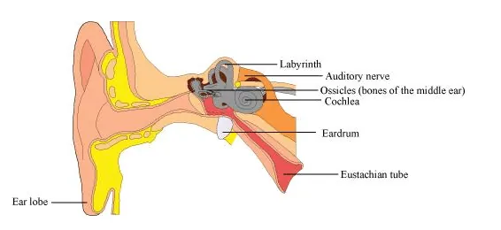
(C) Ear: Ear is the sense organ for hearing and equilibrium. It consists of three portions – external ear, middle ear, and internal ear.
1. External ear:
It consists of pinna, external auditory meatus, and a tympanic membrane.
(a) Pinna is a sensitive structure that collects and directs the vibrations into the ear to produce sound.
(b) External auditory meatus is a tubular passage supported by cartilage in external ear.
(c) Tympanic membrane is a thin membrane that lies close to the auditory canal. It separates the middle ear from external ear.
2. Middle ear:
It is an air-filled tympanic cavity that is connected with pharynx through eustachian tube. Eustachian tube helps to equalize air pressure in both sides of tympanic membrane. The middle ear contains a flexible chain of three middle bones called ear ossicles. The three ear ossicles are malleus, incus, and stapes that are attached to each other.
3. Internal ear:
It is also known as labyrinth. Labyrinth is divided into bony labyrinth and a membranous labyrinth. Bony labyrinth is filled with perilymph while membranous labyrinth is filled with endolymph. Membranous labyrinth is divided into 2 parts.
(a) Vestibular apparatus Vestibular apparatus is a central sac-like part that is divided into utriculus and sacculus. A special group of sensory cells called macula are present in sacculus and utriculus.
Vestibular apparatus also contains three semi-circular canals. The lower end of each semi-circular canal contains a projecting ridge called crista ampularis. Each ampulla has a group of sensory cells called crista. Crista and macula are responsible for maintaining the balance of body and posture.
(b) Cochlea:
Cochlea is a long and coiled outgrowth of sacculus. It is the main hearing organ. Cochlea consists of three membranes. The organ of corti, a hearing organ, is located on the basilar membrane that has hair cells.
Question 2: Compare the following:
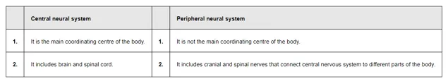
(a) Central neural system (CNS) and Peripheral neural system (PNS)
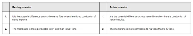
(b) Resting potential and action potential
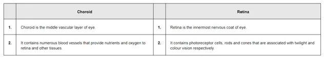
(c) Choroid and retina
Solution. (a) Central neural system (CNS) and Peripheral neural system (PNS)
(b) Resting potential and action potential
(c) Choroid and retina
Question 3: Explain the following processes:
(a) Polarisation of the membrane of a nerve fibre
(b) Depolarisation of the membrane of a nerve fibre
(c) Conduction of a nerve impulse along a nerve fibre
(d) Transmission of a nerve impulse across a chemical synapse
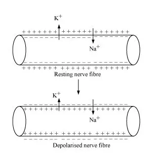
Solution. (a) Polarisation of the membrane of a nerve fibre
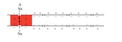
move faster from inside to outside as compared to sodium ions. Therefore, the membrane becomes positively charged outside and negatively charged inside. This is known as polarization of membrane or polarized nerve.
(b) Depolarisation of the membrane of a nerve fibre
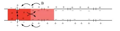
When an electrical stimulus is given to a nerve fibre, an action potential is generated. The membrane becomes permeable to sodium ions than to potassium ions. This results into positive charge inside and negative charge outside the nerve fibre. Hence, the membrane is said to be depolarized.
(c) Conduction of a nerve impulse along a nerve fibre
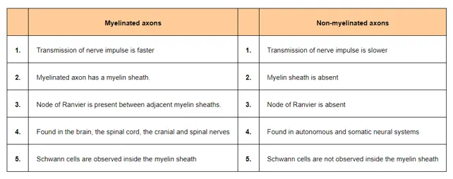
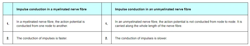
There are two types of nerve fibres – myelinated and non-myelinated. In myelinated nerve fibre, the action potential is conducted from node to node in jumping manner. This is because the myelinated nerve fibre is coated with myelin sheath. The myelin sheath is impermeable to ions. As a result, the ionic exchange and depolarisation of nerve fibre is not possible along the whole length of nerve fibre. It takes place only at some point, known as nodes of Ranvier, whereas in non-myelinated nerve fibre, the ionic exchange and depolarization of nerve fibre takes place along the whole length of the nerve fibre. Because of this ionic exchange, the depolarized area becomes repolarised and the next polarized area becomes depolarized.
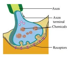
(d) Transmission of a nerve impulse across a chemical synapse
Synapse is a small gap that occurs between the last portion of the axon of one neuron and the dendrite of next neuron. When an impulse reaches at the end plate of axon, vesicles consisting of chemical substance or neurotransmitter, such as acetylcholine, fuse with the plasma membrane. This chemical moves across the cleft and attaches to chemo-receptors present on the membrane of the dendrite of next neuron. This binding of chemical with chemo-receptors leads to the depolarization of membrane and generates a nerve impulse across nerve fibre.
The chemical, acetylcholine, is inactivated by enzyme acetylcholinestrase. The enzyme is present in the post synaptic membrane of the dendrite.
It hydrolyses acetylcholine and this allows the membrane to repolarise.
Question 4: Draw labelled diagrams of the following:
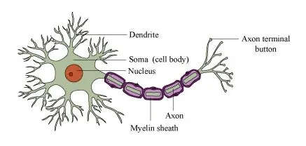
(a) Neuron
(b) Brain
(c) Eye
(d) Ear
Solution. (a) Neuron
(b) Brain
(c) Eye
(d) Ear
Question 5: Write short notes on the following:
(a) Neural coordination
(b) Forebrain
(c) Midbrain
(d) Hindbrain
(e) Retina
(f) Ear ossicles
(g) Cochlea
(h) Organ of Corti
(i) Synapse
Solution. (a) Neural coordination
The neural system provides rapid coordination among the organs of the body. This coordination is in the form of electric impulses and is quick and short lived. All the physiological processes in the body are closed linked and dependent upon each other. For example, during exercise, our body requires more oxygen and food. Hence, the breathing rate increases automatically and the heart beats faster. This leads to a faster supply of oxygenated blood to the muscles. Moreover, the cellular functions require regulation continuously. These functions are carried out by the hormones. Hence, the neural system along with the endocrine system control and coordinate the physiological processes.
(b) Forebrain
It is the main thinking part of the brain. It consists of cerebrum, thalamus, and hypothalamus.
(i) Cerebrum:
Cerebrum is the largest part of the brain and constitutes about four-fifth of its weight. Cerebrum is divided into two cerebral hemispheres by a deep longitudinal cerebral fissure. These hemispheres are joined by a tract of nerve fibres known as corpus callosum. The cerebral hemispheres are covered by a layer of cells known as cerebral cortex or grey matter. Cerebrum has sensory regions known as association areas that receive sensory impulses from various receptors as well as from motor regions that control the movement of various muscles. The innermost part of cerebrum gives an opaque white appearance to the layer and is known as the white matter.
(ii) Thalamus:
Thalamus is the main centre of coordination for sensory and motor signalling. It is wrapped by cerebrum.
(iii) Hypothalamus:
It lies at the base of thalamus and contains a number of centres that regulate body temperature and the urge for eating and drinking. Some regions of cerebrum, along with hypothalamus, are involved in the regulation of sexual behaviour and expression of emotional reactions such as excitement, pleasure, fear, etc.
(c) Midbrain
It is located between the thalamus region of the forebrain and pons region of hindbrain. The dorsal surface of midbrain consists of superior and inferior corpora bigemina and four round lobes called corpora quadrigemina. A canal known as cerebral aqueduct passes through the midbrain. Midbrain is concerned with the sense of sight and hearing.
(d) Hindbrain
It consists of three regions – pons, cerebellum, and medulla oblongata.
(i) Pons is a band of nerve fibres that lies between medulla oblongata and midbrain. It connects the lateral parts of cerebellar hemisphere together.
(ii) Cerebellum is a large and well developed part of hindbrain. It is located below the posterior sides of cerebral hemispheres and above the medulla oblongata. It is responsible for maintaining posture and equilibrium of the body.
(iii) Medulla oblongata is the posterior and simplest part of the brain. It is located beneath the cerebellum. Its lower end extends in the form of spinal cord and leaves the skull through foramen magnum.
(e) Retina
Retina is the innermost layer. It contains three layers of cells – inner ganglion cells, middle bipolar cells, and outermost photoreceptor cells. The receptor cells present in the retina are of two types – rod cells and cone cells.
(i) Rod cells –The rods contain rhodopsin pigment (visual purple), which is highly sensitive to dim light. It is responsible for twilight vision.
(ii) Cone cells –The cones contain iodopsin pigment (visual violet) and are highly sensitive to high intensity light. They are responsible for daylight and colour visions.
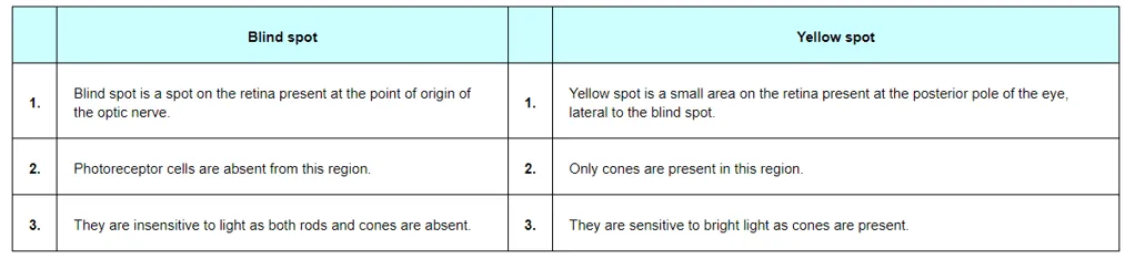
The innermost ganglionic cells give rise to optic nerve fibre that forms optic nerve in each eye and is connected with the brain. In this region, the photoreceptor cells are absent. Hence, it is known as the blind spot. At the posterior part, lateral to blind spot, there is a pigmented spot called macula lutea. This spot has a shallow depression at its middle known as fovea. Fovea has only cone cells. They are devoid of rod cells. Hence, it is the place of most distinct vision.
(f) Ear ossicles
The middle ear contains a flexible chain of three middle bones called ear ossicles. The three ear ossicles are as follows.
(i) Malleus
(ii) Incus
(iii) Stapes
The malleus is attached to tympanic membrane on one side and to incus on the other side. The incus is connected with stapes. Stapes, in turn, are attached with an oval membrane, fenestra ovalis, of internal ear. The ear ossicles act as a lever that transmits sound waves from external ear to internal ear.
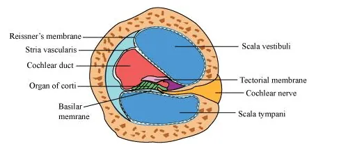
(g) Cochlea
Cochlea is a long, coiled outgrowth of sacculus. It is the main hearing organ. The cochlea forms three chambers.
(i) Upper − scala vestibule
(ii) Middle − scala media
(iii) Lower − scale tympani
The floor of the scala media is basilar membrane while its roof is Reissner’s membrane. Reissner’s membrane gives out a projection called tectorial membrane. The organ of corti, a hearing organ, is located on the basilar membrane. Organ of corti contains receptor hair cells. The upper scala vestibule and lower scala tympani contain perilymph.
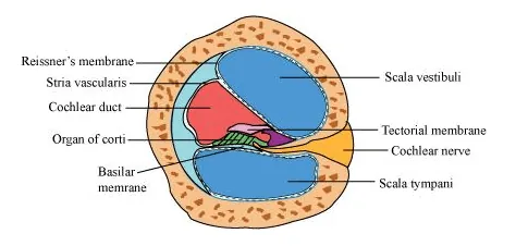
(h) Organ of corti
Organ of corti is the hearing organ. It is located on the basilar membrane that contains hair cells. Hair cells act as auditory receptors. They are present on the internal side of organ of corti.
(i) Synapse
Synapse is a junction between the axon terminal of one neuron and the dendrite of next neuron. It is separated by a small gap known as synaptic cleft.
There are two types of synapses.
(a) Electrical synapse
(b) Chemical synapse
In electrical synapses, the pre and post synaptic neurons lie in close proximity to each other. Hence, the impulse can move directly from one neuron to another across the synapse. This represents a faster method of impulse transmission.
In chemical synapses, the pre and post synaptic neurons are not in close proximity. They are separated by a synaptic cleft. The transmission of nerve impulses is carried out by chemicals such as neurotransmitters.
Question 6:
Give a brief account of:
(a) Mechanism of synaptic transmission
(b) Mechanism of vision
(c) Mechanism of hearing
Solution. (a) Mechanism of synaptic transmission
Synapse is a junction between two neurons. It is present between the axon terminal of one neuron and the dendrite of next neuron separated by a cleft.
There are two ways of synaptic transmission.
(1) Chemical transmission
(2) Electrical transmission
1. Chemical transmission – When a nerve impulse reaches the end plate of axon, it releases a neurotransmitter (acetylcholine) across the synaptic cleft. This chemical is synthesized in cell body of the neuron and is transported to the axon terminal. The acetylcholine diffuses across the cleft and binds to the receptors present on the membrane of next neuron. This causes depolarization of membrane and initiates an action potential.
2. Electrical transmission – In this type of transmission, an electric current is formed in the neuron. This electric current generates an action potential and leads to transmission of nerve impulse across the nerve fibre. This represents a faster method of nerve conduction than the chemical method of transmission.
(b) Mechanism of vision
Retina is the innermost layer of eye. It contains three layers of cells – inner ganglion cells, middle bipolar cells, and outermost photoreceptor cells. A photoreceptor cell is composed of a protein called opsin and an aldehyde of vitamin A called retinal. When light rays are focused on the retina through cornea, it leads to the dissociation of retinal from opsin protein. This changes the structure of opsin. As the structure of opsin changes, the permeability of membrane changes, generating a potential difference in the cells. This generates an action potential in the ganglionic cells and is transmitted to the visual cortex of the brain via optic nerves. In the cortex region of brain, the impulses are analysed and image is formed on the retina.
(c) Mechanism of hearing
The pinna of the external region collects the sound waves and directs it towards ear drum or external auditory canal. These waves strike the tympanic membrane and vibrations are created. Then, these vibrations are transmitted to the oval window, fenestra ovalis, through three ear ossicles, named as malleus, incus, and stapes. These ear ossicles act as lever and transmit the sound waves to internal ear. These vibrations from fenestra ovalis are transmitted into cochlear fluid. This generates sound waves in the lymph. The formation of waves generates a ripple in the basilar membrane. This movement bends the sensory hair cells present on the organ of corti against tectorial membrane. As a result of this, sound waves are converted into nerve impulses. These impulses are then carried to auditory cortex of brain via auditory nerves. In cerebral cortex of brain, the impulses are analysed and sound is recognized.
Question 7: Answer briefly:
(a) How do you perceive the colour of an object?
(b) Which part of our body helps us in maintaining the body balance?
(c) How does the eye regulate the amount of light that falls on the retina?
Solution. (a) Photoreceptors are cells that are sensitive to light. They are of two types – rods and cones. These are present in the retina. Cones help in distinguishing colours. There are three types of cone cells – those responding to green light, those responding to blue light, and those responding to red light. These cells are stimulated by different lights, from different sources. The combinations of the signals generated help us see the different colours.
(b) Vestibular apparatus is located in the internal ear, above the cochlea and helps in maintaining body balance. Crista and macula are the sensory spots of the vestibular apparatus controlling dynamic equilibrium.
(c) Pupil is the small aperture in the iris that regulates the amount of light entering the eye. Cornea, aqueous humour, lens, and vitreous humour act together and refract light rays, focussing them onto the photoreceptor cells of the retina.
Question 8:
Explain the following:
(a) Role of Na+ in the generation of action potential.
(b) Mechanism of generation of light-induced impulse in the retina.
(c) Mechanism through which a sound produces a nerve impulse in the inner ear.
Solution. (a) Sodium ions play an important role in the generation of action potential. When a nerve fibre is stimulated, the membrane potential decreases. The membrane becomes more permeable to $\mathrm{Na}^{+}$ions than to $\mathrm{K}^{+}$ions. As a result, $\mathrm{Na}^{+}$diffuses from the outside to the inside of the membrane. This causes the inside of the membrane to become positivelycharged, while the outer membrane gains a negatively charge. This reversal of polarity across the membrane is known as depolarisation. The rapid inflow of $\mathrm{Na}^{+}$ions causes the membrane potential to increase, thereby generating an action potential.
(b) Retina is the innermost layer of the eye. It contains three layers of cells - inner ganglion cells, middle bipolar cells, and outermost photoreceptor cells. Photoreceptor cells are composed of a protein called opsin and an aldehyde of vitamin A called retinal. When light rays are focused on the retina through the cornea, retinal gets dissociated from opsin. As a result, the structure of opsin gets changed. This in turn causes the permeability of the membrane to change, thereby generating a potential difference in the cells. Consequently, an action potential is generated in the ganglion cells and is transmitted to the visual cortex of the brain via the optic nerves. In the cortex region of the brain, the impulses are analysed and the image is formed on the retina
(c) The pinna of the external ear collects the sound waves and directs them to the tympanic membrane (ear drum) via the external auditory canal. The ear drum then vibrates the sound waves and conducts them to the internal ear through the ear ossicles. The ear ossicles increase the intensity of the sound waves. These vibrating sound waves are conducted through the oval window to the fluid in the cochlea. Consequently, a movement is created in the lymph. This movement produces vibrations in the basilar membrane, which in turn stimulate the auditory hair cells. These cells generate a nerve impulse, conducting it to the auditory cortex of the brain via afferent fibres. The auditory cortex region interprets the nerve impulse and sound is recognised.
Question 9: Differentiate between:
(a) Myelinated and non-myelinated axons
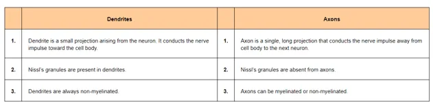
(b) Dendrites and axons
(c) Rods and cones
(d) Thalamus and Hypothalamus
(e) Cerebrum and Cerebellum
Solution. (a) Myelinated and non-myelinated axons
(b) Dendrites and axons
(c) Rods and cones
(d) Thalamus and Hypothalamus
(e) Cerebrum and Cerebellum
Question 10: Answer the following:
(a) Which part of the ear determines the pitch of a sound?
(b) Which part of the human brain is the most developed?
(c) Which part of our central neural system acts as a master clock?
Solution. (a) Cochlea determines the pitch of a sound.
(b) Forebrain is largest and the most developed part of the human brain.
(c) Hypothalamus acts as a master clock in the human body.
Question 11: The region of the vertebrate eye, where the optic nerve passes out of the retina, is called the
(a) fovea
(b) iris
(c) blind spot
(d) optic chaisma
Solution. Answer: (c) Blind spot
Blind spot is the part where the optic nerve passes out of the retina. Photoreceptors are absent from this region.
Question 12: Distinguish between:
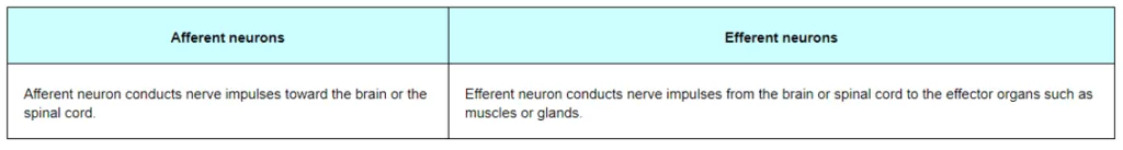
(a) afferent neurons and efferent neurons
(b) impulse conduction in a myelinated nerve fibre and unmyelinated nerve fibre
(c) aqueous humor and vitreous humorv
(d) blind spot and yellow spot
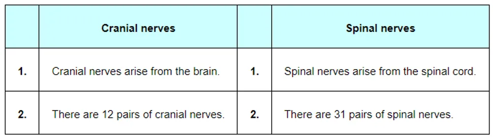
(f) cranial nerves and spinal nerves.
Solution. (a) Afferent neurons and efferent neurons
(b) Impulse conduction in a myelinated nerve fibre and an unmyelinated nerve fibre
c) Aqueous humour and vitreous humour
(d) Blind spot and yellow spot
(e) Cranial nerves and spinal nerves
Also Read,
Class 11 Chemistry Notes.
Class 11 Biology Book Chapterwise.
Class 11 Biology Exemplar Chapterwise.
If you have any Confusion related to NCERT Solutions for Class 11 Biology chapter 21 Neural Control and Coordination PDF then feel free to ask in the comments section down below.
To watch Free Learning Videos on Class 11 Biology by Kota’s top Doctor’s Faculties Install the eSaral App