Parts of a Typical Angiosperm Ovule — Labelled Diagram, Structure & Functions
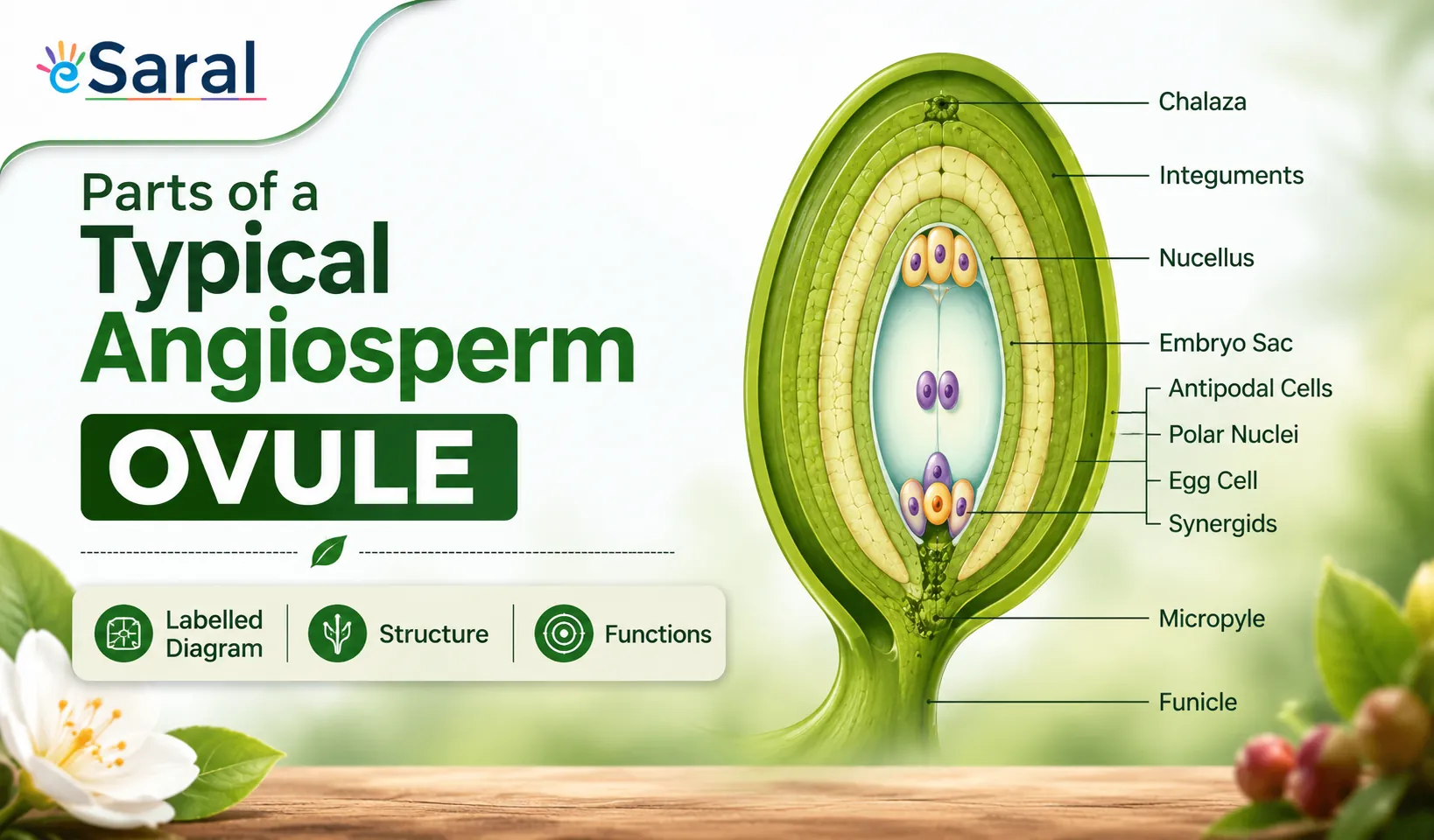


With a neat, labelled diagram, describe the parts of a typical angiosperm ovule.
An ovule is a female megasporangium where the formation of megaspores takes place.
The various parts of an ovule are –
(1) Funiculus – It is a stalk-like structure which represents the point of attachment of the ovule to the placenta of the ovary.
(2) Hilum – It is the point where the body of the ovule is attached to the funiculus.
(3) Integuments –They are the outer layers surrounding the ovule that provide protection to the developing embryo.
(4) Micropyle – It is a narrow pore formed by the projection of integuments. It marks the point where the pollen tube enters the ovule at the time of fertilization.
(5) Nucellus – It is a mass of parenchymatous tissue surrounded by the integuments from the outside. The nucellus provides nutrition to the developing embryo. The embryo sac is located inside the nucellus.
(6) Chalazal – It is the based swollen part of the nucellus from where the integuments originate.
What Is an Angiosperm Ovule?
The ovule is a fundamental structure in the sexual reproduction of flowering plants (angiosperms). It is the female megasporangium — the site where the megaspore mother cell undergoes meiosis to produce megaspores, one of which develops into the female gametophyte (embryo sac).
After fertilisation, the ovule matures into a seed, and the ovary wall develops into the fruit. Understanding the ovule is, therefore, the starting point for understanding how angiosperms reproduce, how seeds are formed, and how genetic material is passed from one generation to the next.
This topic appears in Chapter 2: Sexual Reproduction in Flowering Plants of Class 12 Biology (NCERT), and carries consistent weightage in NEET UG and board examinations. For NCERT-aligned solutions and detailed chapter notes, you can explore NCERT Solutions Class 12 and NCERT Books Class 12 on eSaral.
What Are the Parts of a Typical Angiosperm Ovule?
A typical anatropous angiosperm ovule has six main structural components. Each part has a distinct position and a specific role in protecting the developing embryo and ensuring successful fertilisation.
1. Funiculus
The funiculus is the stalk-like structure that physically connects the ovule to the placenta of the ovary wall. It acts as the supply line, conducting nutrients and water from the parent plant into the ovule. In an anatropous ovule (the most common type in angiosperms), the funiculus is fused along the body of the ovule, forming a ridge called the raphe.
2. Hilum
The hilum is the exact point where the body of the ovule merges with the funiculus. It is the equivalent of a navel — marking the attachment scar. In seeds, the hilum remains visible as a scar on the seed coat (testa), which is why it is easy to identify in practical examinations.
3. Integuments
Integuments are the protective outer layers that envelop the nucellus. Most angiosperms have two integuments (bitegmic ovules) — an outer integument and an inner integument — though some families have only one (unitegmic). After fertilisation, the integuments harden and differentiate into the seed coat (testa and tegmen), protecting the embryo during dormancy and germination.
4. Micropyle
The micropyle is a narrow, pore-like opening formed where the integuments do not completely close at the tip of the ovule.
It serves a dual purpose:
- It is the entry point for the pollen tube during fertilisation.
- In seeds, it allows water absorption during germination (imbibition).
In NEET, a common question asks which structure guides the pollen tube into the ovule. The answer is always the micropyle — not the chalaza or hilum. Remember: micro = small, pyle = gate. It is the "small gate" through which the pollen tube enters.
5. Nucellus
The nucellus is the central mass of parenchymatous (thin-walled, storage) tissue enclosed within the integuments. It is the nutritive tissue of the ovule and plays two key roles:
- It nourishes the developing embryo sac and later the embryo.
- It houses the embryo sac (female gametophyte), which is embedded within it.
In some species, nucellus tissue persists into the seed as perisperm (e.g., black pepper, cardamom), which is a modified form of nutritive tissue distinct from endosperm.
6. Chalaza
The chalaza is the basal, swollen region of the nucellus — the end opposite the micropyle. It is the point from which the integuments originate. The chalaza also plays a role in transporting nutrients from the funiculus into the nucellus. The region of the ovule between the chalaza and the embryo sac is sometimes called the chalazal end and is important in describing the polarity of the embryo sac.
Labelled Diagram of an Angiosperm Ovule

What Is the Function of Each Part of the Ovule?
The table below provides a quick-reference summary of all six parts, their position, and their primary function — ideal for last-minute NEET revision.
| Part | Position in Ovule | Primary Function |
|---|---|---|
| Funiculus | Stalk connecting ovule to placenta | Attachment + nutrient/water conduction |
| Hilum | Junction of funiculus and ovule body | Marks point of attachment; visible as seed scar |
| Outer Integument | Outermost protective layer | Develops into testa (outer seed coat) |
| Inner Integument | Layer inside outer integument | Develops into tegmen (inner seed coat) |
| Micropyle | Pore at the tip, between integuments | Entry of pollen tube; water absorption in seeds |
| Nucellus | Central parenchymatous tissue | Nourishes embryo sac and embryo; may form perisperm |
| Chalaza | Basal swollen region of nucellus | Origin point of integuments; nutrient transfer |
| Embryo Sac | Located within nucellus | Female gametophyte — site of fertilisation |
Students at eSaral's NEET batches — including those who scored 650+ in NEET 2024 — consistently use the mnemonic "Funny Hippos In My New Canteen" (Funiculus → Hilum → Integuments → Micropyle → Nucellus → Chalaza) to recall all six parts in the correct order from stalk to base.
Types of Ovules Found in Angiosperms
Not all ovules have the same orientation relative to the funiculus. The NCERT Class 12 Biology textbook describes six main types, of which anatropous is the most common in angiosperms.
| Type | Description | Example |
|---|---|---|
| Orthotropous (Atropous) | Ovule erect; micropyle faces upward, away from funiculus | Polygonum, Piper |
| Anatropous | Ovule inverted 180°; micropyle faces downward near hilum | Most angiosperms (e.g., sunflower) |
| Campylotropous | Ovule curved so embryo sac is bent | Legumes (e.g., gram) |
| Hemianatropous | Ovule at right angles to funiculus | Ranunculus |
| Amphitropous | Both ovule and embryo sac are curved | Alismaceae |
| Circinotropous | Funiculus is very long, coiled around ovule | Opuntia |
How Does the Ovule Relate to the Embryo Sac?
The ovule and the embryo sac are closely linked but are not the same structure.
- The ovule is the megasporangium — the entire female reproductive unit.
- The embryo sac (female gametophyte) is a structure inside the ovule, formed within the nucellus from a functional megaspore.
Development Sequence Inside the Ovule
- A megaspore mother cell (MMC) inside the nucellus undergoes meiosis and produces four megaspores.
- Three megaspores degenerate; one functional megaspore survives.
- The functional megaspore undergoes three rounds of mitosis and forms an eight-nucleate embryo sac.
- The mature embryo sac contains an egg cell, two synergids, three antipodal cells, and two polar nuclei (central cell).
After double fertilisation:
- Egg cell + male gamete → zygote → embryo
- Central cell (polar nuclei) + male gamete → primary endosperm nucleus → endosperm
- Ovule as a whole → seed
NEET & Board Exam Tips for This Topic
NEET UG (conducted by NTA) regularly features 1–2 questions directly from the ovule structure and embryo sac topic.
High-Frequency NEET Question Areas
- Function of micropyle — pollen tube entry and water imbibition
- Number of cells in a mature embryo sac: 7 cells, 8 nuclei
- Perisperm vs endosperm — perisperm from nucellus; endosperm from triple fusion
- Anatropous ovule — most common type; be able to draw and label it
- Integuments form the seed coat — outer = testa, inner = tegmen
Board Exam (CBSE Class 12) Tips
- A diagram question on the ovule is worth 3–5 marks in board exams.
- Always label: funiculus, hilum, micropyle, outer integument, inner integument, nucellus, embryo sac, chalaza, and raphe.
- Write answers in point form with brief explanations for each part.
Frequently Asked Questions
Find answers to common questions.
What is the function of the micropyle in an ovule?
The micropyle is a small pore formed by the incomplete closure of the integuments at the tip of the ovule. Its primary function is to allow the pollen tube to enter the ovule during fertilisation. In seeds, the micropyle also permits water to enter during germination, a process called imbibition that activates seed metabolism.
How many parts does a typical angiosperm ovule have?
A typical angiosperm ovule has six main structural parts: funiculus, hilum, integuments (usually two — outer and inner), micropyle, nucellus, and chalaza. Additionally, the embryo sac (female gametophyte) is housed inside the nucellus and is considered a part of the ovule's functional anatomy in most Class 12 and NEET-level descriptions.
What is an angiosperm ovule?
An angiosperm ovule is the female megasporangium located inside the ovary of a flowering plant. It is the structure where megaspores are produced by meiosis, and where the female gametophyte (embryo sac) develops. After fertilisation, the entire ovule matures into a seed, making it central to sexual reproduction in angiosperms.
What is the chalaza in an ovule and why is it important?
The chalaza is the basal, swollen region of the nucellus located at the end opposite the micropyle. It is the point from which the integuments originate and grow upward to envelope the nucellus. The chalaza also serves as a nutrient transfer zone, channelling food materials from the funiculus into the nucellus. In some seeds, the chalaza remains visible as a distinct scar
What type of ovule is most common in angiosperms?
The anatropous ovule is the most common type in angiosperms. In an anatropous ovule, the body of the ovule is completely inverted (turned 180°) so that the micropyle faces downward toward the hilum and funiculus. The funiculus fuses with the body of the ovule to form the raphe. Sunflower and most common crop plants have anatropous ovules.
What is the difference between nucellus and integuments?
The nucellus is the central mass of parenchymatous tissue inside the ovule that nourishes the embryo sac and developing embryo. The integuments are the outer protective layers surrounding the nucellus. After fertilisation, the nucellus may become perisperm, while the integuments differentiate into the seed coat — the outer integument into the testa and the inner integument into the tegmen.